
Key Takeaways
- Accurate diagnosis hinges on a blend of history, clinical rating scales, and targeted imaging.
- Video analysis and sensor‑based assessments boost objectivity.
- DaTscans, MRI, and PET each offer distinct clues about underlying brain changes.
- Choosing the right test depends on symptom severity, resource availability, and patient comorbidities.
Dyskinesia is a movement disorder marked by involuntary, repetitive motions, frequently emerging as a side‑effect of dopaminergic therapy in Parkinson’s disease. Because the movements can mimic tremor or chorea, clinicians need a systematic approach to separate drug‑induced dyskinesia from other motor disturbances.
Understanding the Clinical Context
Most patients who develop dyskinesia are living with Parkinson's disease, a progressive neurodegenerative condition characterized by loss of dopaminergic neurons in the substantia nigra. When levodopa is introduced, the brain’s fluctuating dopamine levels can trigger the abnormal movements we call levodopa‑induced dyskinesia.
Core Clinical Assessment
A thorough clinical interview remains the backbone of any diagnosis. Key questions include medication timing, dose changes, and the exact onset of involuntary movements. Physical examination should document the distribution (limb vs. axial), quality (ballistic, choreic, or dystonic), and triggering factors (e.g., peak dose periods).
Standardized Rating Scales
Two rating tools dominate practice:
- Unified Parkinson's Disease Rating Scale (UPDRS), specifically Part IV, quantifies motor complications including dyskinesia. Scores range from 0 (none) to 4 (severe), offering a numeric anchor for treatment decisions.
- Abnormal Involuntary Movement Scale (AIMS) was originally designed for antipsychotic‑induced movements, but many neurologists adapt it for dyskinesia because it captures facial, oral, and trunk involvement in a single pass.
Both scales are quick (<5min) and can be repeated at each clinic visit to track progression.
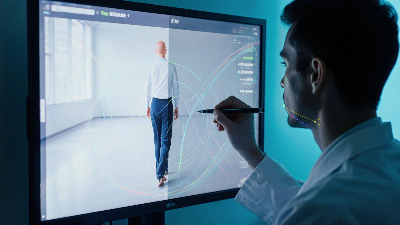
Instrumented Video Analysis
High‑definition video recorded at the patient’s peak‑dose window, followed by frame‑by‑frame review, adds visual proof. Modern platforms allow clinicians to overlay motion‑tracking markers, calculating amplitude (degrees) and frequency (Hz). This objective data is especially useful for research trials and for discussing medication adjustments with patients.
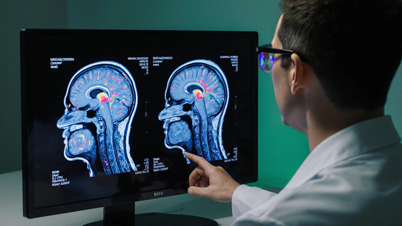
Neuroimaging Options
Imaging never replaces a solid clinical exam, but it can reveal structural or functional changes that guide therapy.
- DaTscan (Ioflupane SPECT) visualizes dopamine transporter density. In dyskinesia caused by levodopa, the scan often appears normal, helping rule out atypical Parkinsonian syndromes.
- Magnetic Resonance Imaging (MRI), especially with diffusion tensor imaging, can detect microstructural changes in the basal ganglia that correlate with dyskinetic severity.
- Positron Emission Tomography (PET) using fluorodopa or FDG tracers maps metabolic activity. Elevated metabolic rates in the putamen are a hallmark of peak‑dose dyskinesia.
Choosing between them depends on cost, local availability, and the clinical question: rule‑out diagnosis (DaTscan), structural insight (MRI), or functional metabolism (PET).
Laboratory & Biomarker Testing
While no blood test definitively diagnoses dyskinesia, a few ancillary studies can sharpen the picture.
- Plasma levodopa levels measured at peak and trough help confirm whether dose timing aligns with symptom spikes.
- Genetic panels targeting GCH1, TH, and SNCA mutations are useful when dyskinesia appears unusually early or in families with hereditary Parkinsonism.
Decision Flowchart
Below is a quick mental checklist clinicians can run through during a consult:
- Confirm medication regimen and timing.
- Apply UPDRS PartIV and AIMS; score severity.
- If scores indicate moderate‑to‑severe dyskinesia, schedule video capture at peak dose.
- Order DaTscan if differential diagnosis is uncertain; otherwise proceed to MRI for structural detail.
- Reserve PET for atypical cases or when considering deep‑brain stimulation.
- Review levodopa plasma levels and consider genetic testing if onset <40years.
Comparison of Diagnostic Tools
| Tool | Sensitivity | Specificity | Invasiveness | Typical Cost (USD) | Availability |
|---|---|---|---|---|---|
| UPDRS PartIV | ≈85% | ≈80% | Non‑invasive | Free (clinic) | Universal |
| AIMS | ≈78% | ≈85% | Non‑invasive | Free (clinic) | Universal |
| Video analysis | ≈92% | ≈88% | Non‑invasive | 200‑400 | Specialized centers |
| DaTscan (SPECT) | ≈90% | ≈95% | Low‑radiation | 1,200‑1,500 | Major hospitals |
| MRI (DTI) | ≈75% | ≈80% | Non‑invasive | 800‑1,200 | Widely available |
| PET (FDG/Fluorodopa) | ≈95% | ≈92% | Radioactive tracer | 2,500‑3,500 | Research hospitals |
Practical Checklist for Clinicians
- Document exact levodopa dose, timing, and formulation.
- Score UPDRSIV and AIMS during each visit.
- Capture a 5‑minute video at the patient’s peak‑dose window.
- Order DaTscan if the diagnosis is unclear; otherwise schedule MRI for baseline structural data.
- Consider PET only when planning advanced therapies like deep‑brain stimulation.
- Review levodopa plasma levels when symptom timing is ambiguous.
- Refer patients <40years old or with a positive family history for genetic testing.
When to Refer to a Specialist
If any of the following appear, send the patient to a movement‑disorder neurologist:
- Severe dyskinesia (UPDRSIV≥3) despite medication adjustments.
- Rapid escalation of symptoms over weeks.
- Co‑existing cognitive decline or psychiatric agitation.
- Unclear differential diagnosis (e.g., atypical Parkinsonism).
Future Directions
Emerging wearables using inertial measurement units (IMUs) promise continuous, home‑based monitoring. Machine‑learning models trained on thousands of video clips are already outperforming human raters in detecting subtle dyskinetic patterns. As these tools become mainstream, the reliance on costly imaging may drop, making diagnosis more accessible in community clinics.
dyskinesia diagnosis now blends seasoned clinical judgment with objective technology. By following a structured assessment pathway, clinicians can differentiate true drug‑induced movements from mimics, adjust therapy promptly, and improve quality of life for patients navigating Parkinson’s disease.
Frequently Asked Questions
What is the first step in diagnosing dyskinesia?
Start with a detailed medication history and a physical exam, then score the Unified Parkinson's Disease Rating Scale PartIV to gauge motor complications.
How reliable is video analysis compared to clinical scales?
When recorded at the patient’s peak‑dose period, video analysis shows sensitivity around 92% and specificity near 88%, often higher than bedside scales alone.
When should I order a DaTscan?
Reserve DaTscan for cases where the clinical picture is ambiguous-especially when you need to exclude atypical Parkinsonian syndromes.
Are there blood tests that confirm dyskinesia?
No single blood test diagnoses dyskinesia, but measuring levodopa plasma concentrations can clarify whether peaks align with involuntary movements.
What role does genetics play in early‑onset dyskinesia?
Mutations in genes such as GCH1, TH, or SNCA can predispose younger patients to severe dyskinesia, so genetic panels are advised for anyone developing symptoms before age40.
How often should I repeat the assessment?
Re‑evaluate every 3‑6months after any medication change, or sooner if patients report a sudden worsening of involuntary movements.


Having gone through the assessment process myself the mix of interview, UPDRS and video checks really helps pinpoint when dyskinesia kicks in. I found that just asking about medication timing can save a lot of guesswork.
Being patient‑centered makes the whole thing feel less clinical.